Introduction
X-Ray beams were identified by the German physicist Will elm Conrad Rontgen in November 1895. He called the ‘revamped sort of flash’ or X-flashes, X for the unknown. their convenience to envision the inward life structures of people was made. Today, imaging with X-flashes is possibly the most regularly utilized demonstrative apparatus with the medicinal calling, and the systems from a basic midsection radiography to a computerized subtraction angiography or machine tomography rely on the utilization of X-flashes.

Properties:
- The principle lands of X-Ray beams, which make them suitable for the purposes of restorative conclusion, are there:
- Proficiencies to infiltrate matter coupled with differential ingestion recognized in diverse materials; and
- Capability to prepare glow and its impact on photographic emulsions.
Principles of X-Ray:
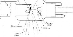
X-beams are transformed in: extraordinarily built glass tube, which fundamentally embodies.
(i) A root for the handling 0: electrons,
(ii) a power origin to quicken the electrons,
(iii) a unlimited electron way,
(iv) a method of cantering the electron bar and
(v) a unit to stop the electrons.
Failure of X-Ray:
There are two principle limits of utilizing accepted X-flashes to test interior structures of the form.
- Firstly, the super-encroachment of the several-dimensional informative content onto a solitary plane makes judgment jumbling and frequently troublesome.
- Also, the photographic picture regularly utilized for making radiographs has a restrained dynamic extend and, consequently, just protests that have substantial differences in X-flash assimilation relative to their surroundings will create sufficient differentiation divergences on the picture to be recognized by the eye.
Conclusion:
Following the time when the CT engineering was improved, quick infrastructures in PC equipment and finder innovation have been witnessed. Advanced CT frameworks get the projection information needed for one tomographic representation in roughly one second and show the recreated picture on a 1024 x 1024 lattice showcase within a few seconds. The representations speak for excellent tomographic maps of the X-flash straight lessening coefficients of the Patient tissues.
